![]()


![]()
Monomethyl auristatin E is an antimitotic agent which inhibits cell division by blocking the polymerisation of tubulin. The linker to the monoclonal antibody is stable in extracellular fluid, but is cleaved by cathepsin once the conjugate has entered a tumor cell, thus activating the antimitotic mechanism. Because of its toxicity, it cannot be used as a drug itself; instead, it is linked to a monoclonal antibody (MAB) which directs it to the cancer cells. In International Nonproprietary Names for MMAE-MAB-conjugates, the name vedotin refers to MMAE plus its linking structure to the antibody. It is a potent antimitotic drug derived from peptides occurring in marine shell-less mollusc Dolabella auricularia called dolastatins which show potent activity in preclinical studies, both in vitro and in vivo, against a range of lymphomas, leukemia and solid tumors. These drugs show potency of up to 200 times that of vinblastine, another antimitotic drug used for Hodgkin lymphoma as well as other types of cancer.
Rabbit
Recombinant Rabbit monoclonal Antibody
1 mg/mL.
ELISA
 | Fig1: Indirect ELISA analysis of MMAE was performed by coating wells of a 96-well plate with 50 μl per well of MMAE-BSA diluted in carbonate/bicarbonate buffer, at a concentration of 1 μg/mL overnight at 4℃. Wells of the plate were washed, blocked with 1%BSA blocking buffer, and incubated with 100 μl per well of MMAE monoclonal antibody serial diluted starting from a concentration of 20ug/ml for 1 hours at room temperature. The plate was washed and incubated with 50 μl per well of an HRP-conjugated goat anti-Rabbit IgG secondary antibody at a dilution of 1:15,000 for one hour at room temperature. Detection was performed using an Ultra TMB Substrate for 10 minutes at room temperature in the dark. The reaction was stopped with sulfuric acid and absorbances were read on a spectrophotometer at 450 nm. |
 | Fig2:Indirect ELISA analysis of MMAE was performed by coating wells of a 96-well plate with 50 μl per well of MMAE-ADC diluted in carbonate/bicarbonate buffer, at a concentration of 1 μg/mL overnight at 4℃. Wells of the plate were washed, blocked with 1%BSA blocking buffer, and incubated with 100 μl per well of MMAE monoclonal antibody serial diluted starting from a concentration of 20ug/ml for 1 hours at room temperature. The plate was washed and incubated with 50 μl per well of an HRP-conjugated goat anti-Rabbit IgG secondary antibody at a dilution of 1:15,000 for one hour at room temperature. Detection was performed using an Ultra TMB Substrate for 10 minutes at room temperature in the dark. The reaction was stopped with sulfuric acid and absorbances were read on a spectrophotometer at 450 nm. |
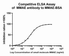 | Fig3:Competitive ELISA analysis of MMAE was performed by coating wells of a 96-well plate with 50 μl per well of MMAE-BSA diluted in carbonate/bicarbonate buffer, at a concentration of 1 μg/mL overnight at 4℃. Wells of the plate were washed, blocked with 1%BSA blocking buffer, and incubated with 100 μl per well of MMAE monoclonal antibody at concentration of 1 μg/mL with serial diluted MMAE starting from a concentration of 10ug/ml for 1 hours at room temperature. The plate was washed and incubated with 50 μl per well of an HRP-conjugated goat anti-Rabbit IgG secondary antibody at a dilution of 1:15,000 for one hour at room temperature. Detection was performed using an Ultra TMB Substrate for 10 minutes at room temperature in the dark. The reaction was stopped with sulfuric acid and absorbances were read on a spectrophotometer at 450 nm. |
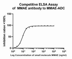 | Fig4:Competitive ELISA analysis of MMAE was performed by coating wells of a 96-well plate with 50 μl per well of MMAE-ADC diluted in carbonate/bicarbonate buffer, at a concentration of 1 μg/mL overnight at 4℃. Wells of the plate were washed, blocked with 1%BSA blocking buffer, and incubated with 100 μl per well of MMAE monoclonal antibody at concentration of 1 μg/mL with serial diluted MMAE starting from a concentration of 10ug/ml for 1 hours at room temperature. The plate was washed and incubated with 50 μl per well of an HRP-conjugated goat anti-Rabbit IgG secondary antibody at a dilution of 1:15,000 for one hour at room temperature. Detection was performed using an Ultra TMB Substrate for 10 minutes at room temperature in the dark. The reaction was stopped with sulfuric acid and absorbances were read on a spectrophotometer at 450 nm. |
Store at +4℃ after thawing. Aliquot store at -20℃. Avoid repeated freeze / thaw cycles.
Note: Application as IHC, only suitable for histochemical staining or fluorescence staining of paraffin-embedded sections. Application as ICC/IF, suitable for histochemical or fluorescent staining of frozen sections, as well as chemical and fluorescent staining at the cellular level.
注意:抗体应用为IHC的,抗体只适合于石蜡切片的组化染色或者荧光染色。
抗体应用为IF/ICC的,抗体适合于冰冻切片的组化染色或者荧光染色,以及细胞水平的化学染色和荧光染色。

艾比玛特医药科技(上海)有限公司
上海市徐汇区桂平路333号聚科生物园区1号楼1-3层
邮箱:market@ab-mart.com
应聘职位:hr@ab-mart.com
订购专线:4006-123-828
销售电话:13162017139(微信同号)
技术支持:15618194176(微信同号)
南方经销商负责:手机13122837132(微信同号)
北方及西南经销商负责:手机13122150513(微信同号)

微信客服
邮箱:market@ab-mart.com
应聘职位:hr@ab-mart.com
订购专线:4006-123-828
销售电话:13162017139(微信同号)
技术支持:15618194176(微信同号)
总机:021-34695901
南方经销商负责:手机13122837132(微信同号)
北方及西南经销商负责:手机13122150513(微信同号)
微信客服
沪ICP备17056956号-2 艾比玛特医药科技(上海)有限公司


